
BRCAura: AI pre-screening for germline BRCA1/2 mutations from routine breast cancer slides
Standard testing criteria based on age, family history, and tumor subtype are estimated to miss about half of germline BRCA1/2 mutation carriers. In a retrospective study of 1,025 patients from six centers in Europe and the US, published in npj Precision Oncology, BRCAura, a deep learning model that reads routine H&E slides, can cut unnecessary sequencing referrals by a third while maintaining sensitivity above 93%.
The gap in BRCA testing
Pathogenic mutations in BRCA1 or BRCA2 are among the most actionable findings in hormone receptor-positive, human epidermal growth factor receptor 2-negative (HR+/HER2−) early breast cancer. They determine eligibility for adjuvant poly(ADP-ribose) polymerase (PARP) inhibitor therapy, inform surgical planning, and trigger cascade genetic testing for at-risk relatives.
Yet genetic testing is usually offered based on age at diagnosis, family history, and tumor subtype. These criteria work well for high-risk families, but population studies estimate that about half of carriers would not be identified by guideline-based testing¹. Testing every patient would close that gap, but cost, sequencing and genetic-counseling capacity, and turnaround times make universal testing difficult in many health systems.
The need is greatest in HR+/HER2− disease. Patients with triple-negative breast cancer are already widely recommended for testing, whereas testing rates in HR+/HER2− patients remain variable and often below guideline targets.
Proof-of-concept studies have explored predicting DNA repair deficiency2 and other molecular markers3 directly from routine histology slides. BRCAura extends this approach to germline BRCA1/2 mutation status in HR+/HER2−early breast cancer, applied at the time of pathological diagnosis with no requirement for family history data.
How BRCAura could fill this gap
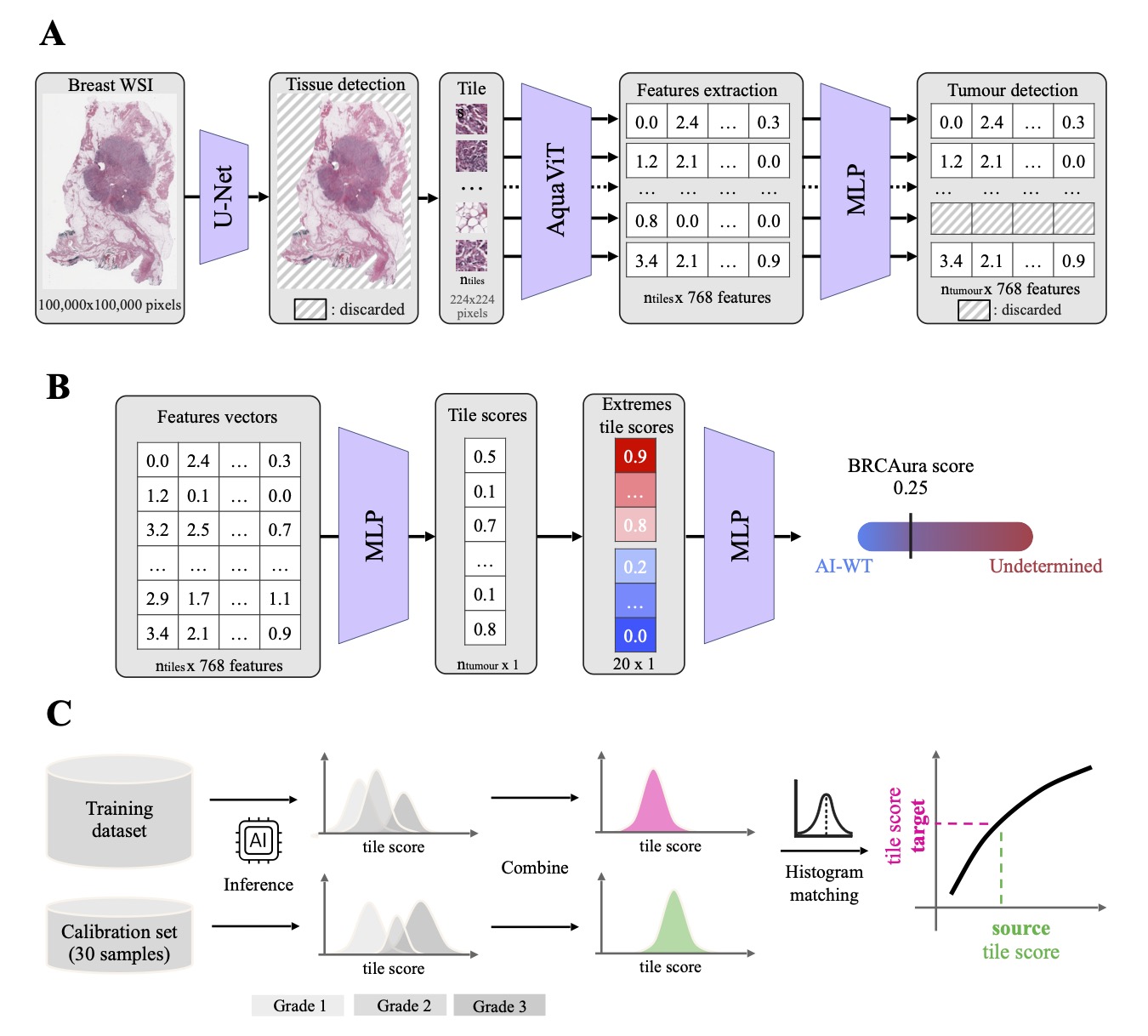
BRCAura analyzes a digitized image of a standard hematoxylin and eosin (H&E) slide that pathologists already prepare for every breast cancer diagnosis. No extra tissue, staining, or family history is needed. The model returns a score from 0 to 1 reflecting how likely the patient is to carry a germline BRCA1/2 mutation (gBRCAm). BRCAura is designed as a rule-out test where slides scoring below a fixed threshold are classified “AI-wild type” (AI-WT) meaning unlikely to carry a mutation. All others are classified “Undetermined”, meaning confirmatory sequencing would still be required. .
The pipeline identifies tissue, and processes tiles it through a state-of-the-art foundation model called AquaViT4, a distilled transformer model built for robustness to scanner and staining variation. A tumor detection step removes non-tumor regions. The multiple instance learning (MIL) model CHOWDER5 then aggregates local predictions into a single slide-level prediction. For each new center in the study, BRCAura was calibrated using 30 slides with known tumor grade, without requiring genetic results. A tile score matching (TSM)⁶ procedure aligned that center’s scores with the training reference so the decision threshold stayed stable across labs, stains, and scanners.
How did BRCAura™ RUO perform in external validation?
To our knowledge, this is the largest external validation of an AI-based gBRCAm classifier in early breast cancer. The dataset comprised 1,025 H&E-stained WSIs from six independent centers across Europe and the United States, collected and analyzed with no involvement from the training institutions. The cohort was assembled deliberately to capture real-world heterogeneity: four scanner platforms (3DHISTECH Pannoramic, Leica Aperio GT450, Roche Ventana DP 600, EasyScan Pro 24), multiple staining protocols, and two specimen types (surgical resections and preoperative biopsies) from centers in Germany, Finland, Spain, and the United States.
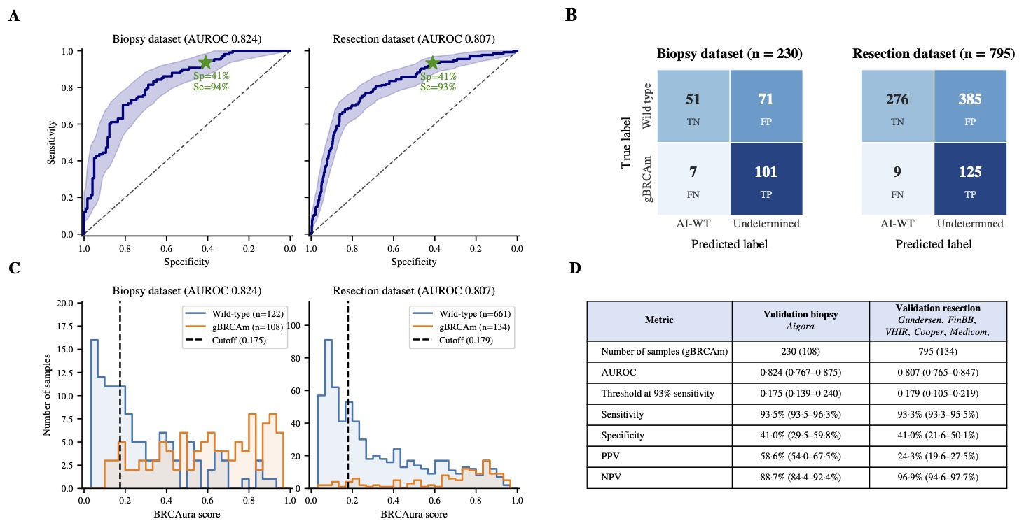
At the operating point targeting at least 93% sensitivity:
- Resection cohort (n = 795, gBRCAm prevalence 16.9%): AUROC 0.807 (95% CI 0.765–0.847). BRCAura ruled out 285 patients (35.8%) , and 96.9% of them were confirmed non-carriers (negative predictive value, NPV). gBRCAm carrier prevalence in the Undetermined group was 24.3%, compared to 16.9% in the unselected cohort.
- Biopsy cohort (n = 230, gBRCAm prevalence 47.0%): 25.2% ruled out; gBRCAm prevalence in the Undetermined group was 58.6%, compared to 47.0% in the unselected cohort. AUROC 0.824 (0.767-0.875), NPV 88.7%.
Analytical repeatability across serial sections stained with H&E and hematoxylin-eosin-saffron (HES) was excellent, with intraclass correlation coefficients of 0.989 and 0.993 respectively. After calibration, performance was consistent across all scanner platforms.
How does BRCAura™ RUO compare with gold standard family-history models like CanRisk?
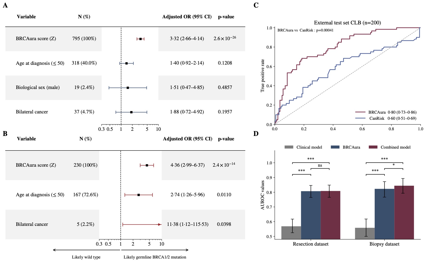
The standard clinical comparator for gBRCAm risk estimation is CanRisk7, a web-based tool used to estimate mutation probability from pedigree information. On the same patients in the Centre Léon Bérard (CLB) external test cohort (n = 200), BRCAura achieved an AUROC of 0.80 (95% CI 0.73-0.86) against 0.60 (0.51-0.69) for CanRisk (DeLong test p=0.00041).
Multivariable logistic regression in the resection cohort identified BRCAura as the only independent predictor of gBRCAm status (odds ratio (OR) 3.32, 95% CI 2.66-4.14; p = 2.5 x 10⁻²⁶). Age at diagnosis, biological sex, and bilaterality were not significant once BRCAura was included in the model. Adding clinical variables to BRCAura did not improve discrimination in the resection cohort: combined AUROC 0.810 versus 0.807 for BRCAura alone.
Unlike CanRisk, BRCAura does not use family history: in this study, its predictions were based on the H&E slide alone.
What does the AI see in the tissue?
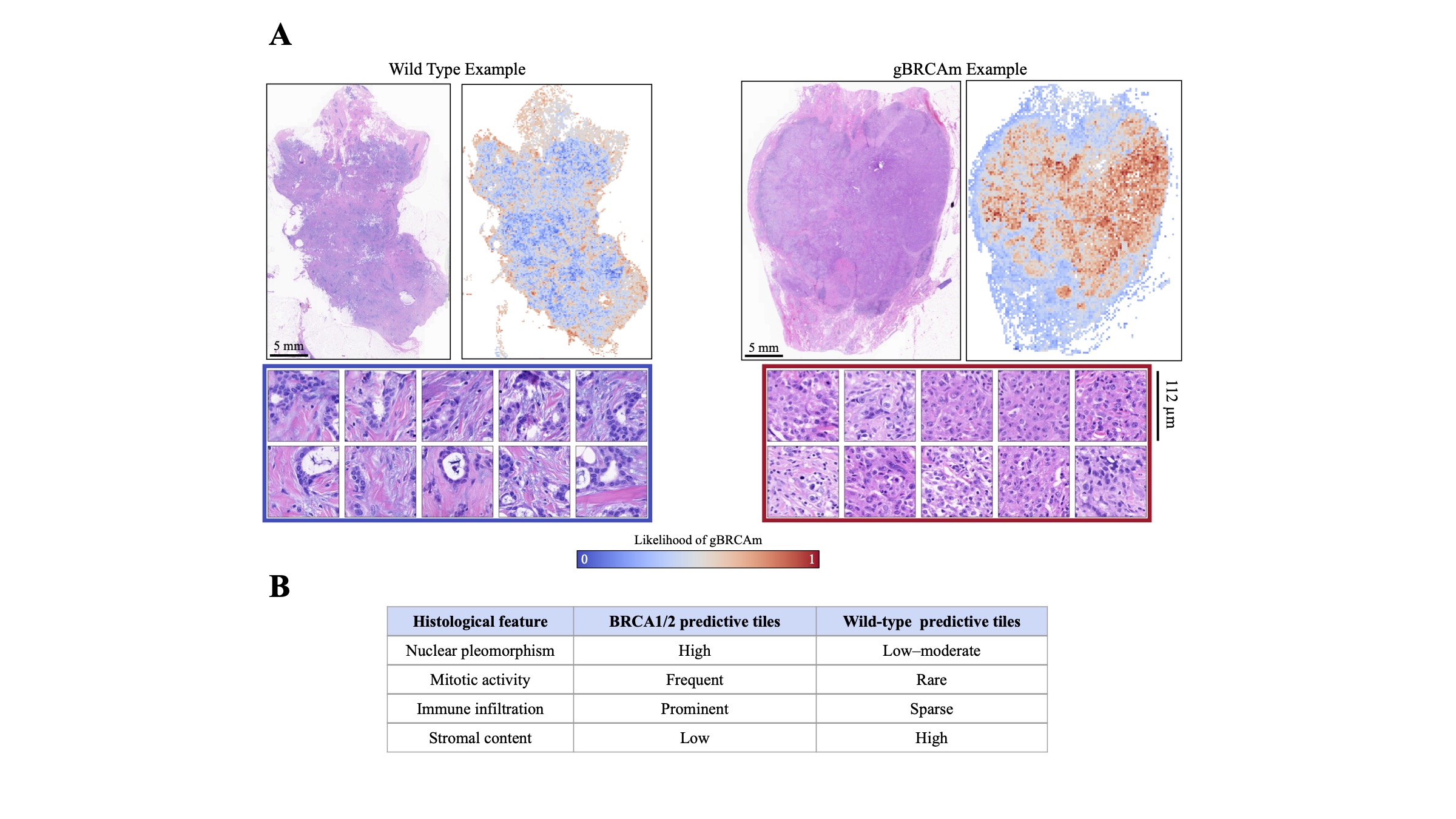
Because BRCAura’s MIL architecture assigns a score to each tile individually, it is possible to map which regions of the slide drive each prediction. High gBRCAm-scoring regions cluster in invasive tumor areas with high cellularity, prominent nucleoli, frequent mitoses, and dense lymphocytic infiltration; low-scoring regions correspond to stromal-rich or hypocellular compartments with abundant fibrous stroma and sparse malignant epithelial cells. These opposing patterns are the established histological features of BRCA-associated breast tumors8.
Quantitative cell counting using an AI-based tumor microenvironment analysis tool9 confirmed significantly higher cancer cell and lymphocyte density in gBRCAm-predictive tiles across all six cohorts (Fisher's exact test, p < 0.001 in each). Wild-type-predictive tiles were richer in fibroblasts. The consistency of these findings across six centers and four scanner platforms gives a mechanistic basis for the predictions and confirms that the model is capturing biological signal rather than acquisition artifacts.
What are the study’s limitations?
The study looked back at existing patient records and slides (a retrospective design), and the patients had mostly been selected for genetic testing because of their risk profile. This means the cohorts contained more carriers than a general breast cancer population would, so some performance figures may differ in broader, unselected populations. Ethnicity data were not available, so performance could not be compared across ethnic groups, and family history was incompletely recorded. Evaluating BRCAura prospectively, in patients enrolled as they are diagnosed, remains an important future step.
What’s next for BRCAura? The Decision Impact Study
Strong validation results show what a test can do, but not whether it can change care. Our new BRCAura Decision Impact Study sets out to answer that question.
A germline BRCA1/2 result can inform eligibility for PARP inhibitor therapy, risk-reducing surgery, surveillance, and testing of family members. This prospective, observational, multicenter study will run across 13 centers in at least six countries, enrolling around 2,600 patients through 2029. It will assess how sharing the BRCAura likelihood category with clinicians influences their decision to recommend germline BRCA1/2 testing. The real-world evidence it generates will help shape how BRCAura could be potentially used in clinical practice.
BRCAura™ RUO is for research use only and is not for use in diagnostic procedures.
FAQ
- Does BRCAura replace genetic testing? No. BRCAura was designed to explore whether AI could help prioritize germline BRCA1/2 testing, making better use of limited sequencing and counseling resources so that more carriers could be found without sequencing every patient. Confirmatory sequencing would still be required, and BRCAura is currently for research use only.
- What types of samples does BRCAura use? Pre-treatment core biopsies (not cytology) or surgical resections from adults with early, non-metastatic, invasive HR+/HER2− breast cancer, scanned at about 0.5 microns per pixel (20x magnification or higher).
- Does BRCAura use family history? No. In this study, its predictions were based on the H&E slide alone.
- Was performance similar for BRCA1 and BRCA2 carriers? Yes. In this study, performance was comparable for both genes.
- Is BRCAura available for clinical use? No. BRCAura is currently available for research use only.
Authors

References
1. Whitworth PW et al. Clinical utility of universal germline genetic testing for patients with breast cancer. JAMA Netw Open 5, e2232787 (2022).
2. Bergstrom EN et al. Deep learning artificial intelligence predicts homologous recombination deficiency and platinum response from histologic slides. J Clin Oncol 42, 3550-3560 (2024). https://doi.org/10.1200/JCO.23.02641
3. Saillard C et al. Validation of MSIntuit as an AI-based pre-screening tool for MSI detection from colorectal cancer histology slides. Nat Commun 14, 6695 (2023).
4. Filiot A et al. Distilling foundation models for robust and efficient models in digital pathology. MICCAI 2025, LNCS vol. 15966, 162-172 (Springer, 2025). https://doi.org/10.1007/978-3-032-04981-0_16
5. Courtiol P et al. Classification and disease localization in histopathology using only global labels: a weakly-supervised approach. Preprint at https://doi.org/10.48550/arXiv.1802.02212 (2020).
6. Pignet A et al. Robust sensitivity control in digital pathology via tile score distribution matching. MICCAI 2025, LNCS, 565-574 (Springer, 2025). https://doi.org/10.1007/978-3-032-04978-0_54
7. Carver T et al. CanRisk tool: a web interface for the prediction of breast and ovarian cancer risk and the likelihood of carrying genetic pathogenic variants. Cancer Epidemiol Biomark Prev 30, 469-473 (2021).
8. Da Silva L & Lakhani SR. Pathology of hereditary breast cancer. Mod Pathol 23, S46-S51 (2010).
9. Adjadj B et al. Towards comprehensive cellular characterisation of H&E slides. Preprint at https://doi.org/10.48550/ARXIV.2508.09926 (2025).