
Fewer slides, faster scoring: a step toward tumor-agnostic IHC AI
presented at ECDP 2026 on tumor-agnostic IHC cell detection and classification
Every new indication for an ADC or targeted therapy can bring a genuinely new biomarker, or an existing one that needs its own scoring criteria for that tumor type. Pathologists then have to learn to score that marker reliably, often on patterns that are genuinely hard to read: heterogeneous expression, subjective thresholds, and high variability between readers. As pipelines expand into more indications at once, this learning curve is becoming a real bottleneck for labs, whether or not they already use AI assisted scoring. And where AI tools do exist, they face a version of the same problem: most are trained on a single marker and a single tumor type, so a new indication typically means training a new model almost from scratch.
At ECDP 2026, the 22nd European Congress on Digital Pathology held from June 17 to 20 in Graz, Austria, the Waiv team presented new research to tackle this challenge. The poster, “Towards Tumor-Agnostic IHC Cell Detection and Classification”, describes a first step towards a model that can generalize across tumor types, investigating both zero-shot transfer and few-shot fine-tuning to new indications and markers as alternative to full retrainings.
The work was authored by Elin Samuelsson, Jean-Eudes Le Douget, Alexandre Filiot, Jérémy Guillon, Benoit Schmauch, David Lin, Cécile Maisin, Kim Vianey, Mina Farag, Corentin Wiscart, Hortense Deslandes, Zoé Vaquette, and Katharina von Loga, with cohort support from Cypath and Cypath-RB.
Why is cell detection the bottleneck in IHC scoring?
Scoring an IHC slide accurately depends on a step that sounds simple but is not: finding every cell in the image and correctly labeling each one as tumor or non-tumor. Errors at this stage can ripple into downstream scores, a HER2 result, a PD-L1 percentage, or any other biomarker readout.
For AI models tackling this task, the usual approach is one model per marker and per tumor type. A lab or biopharma partner working across five indications ends up needing five separate models, each requiring its own large, carefully annotated dataset for its development. As the number of markers and indications grows in step with ADC and targeted therapy development, building a new model for every one of them stops being practical.
How does the model adapt to new tumor types?
The Waiv team built their model around an internal IHC-specific foundation model, used as a frozen feature extractor, paired with a CellViT-based architecture that predicts nuclei locations, separates individual cells using a watershed algorithm, and assigns each cell to a binary tumor class.
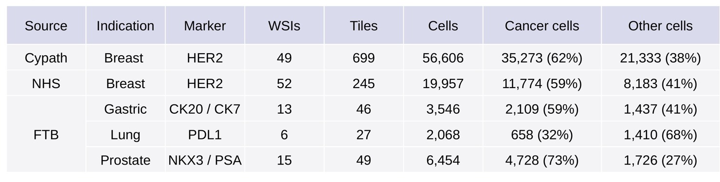
They trained a primary model on a modestly sized breast cancer HER2 cohort from Cypath, then tested it in-domain (same indication and marker) on an independent cohort from the NHS. They also tested it out-of-domain (different indications and markers) on three sub-cohorts from FTB, to measure how well it held up on tumor types and stains it had never seen before.
Then came the more interesting test. Rather than fully retraining the model for each new tumor type, the team performed a joint cross-validation fine-tuning on the FTB cohort, thus training on only 20-30 annotated image tiles from each indication per data split, a small fraction of a typical training set.
Performance was evaluated with a composite metric called panoptic quality, as well as dedicated metrics for each of the cell-level sub-tasks performed by the model: segmentation, detection, and tumor classification.
What did the ECDP 2026 poster demonstrate?
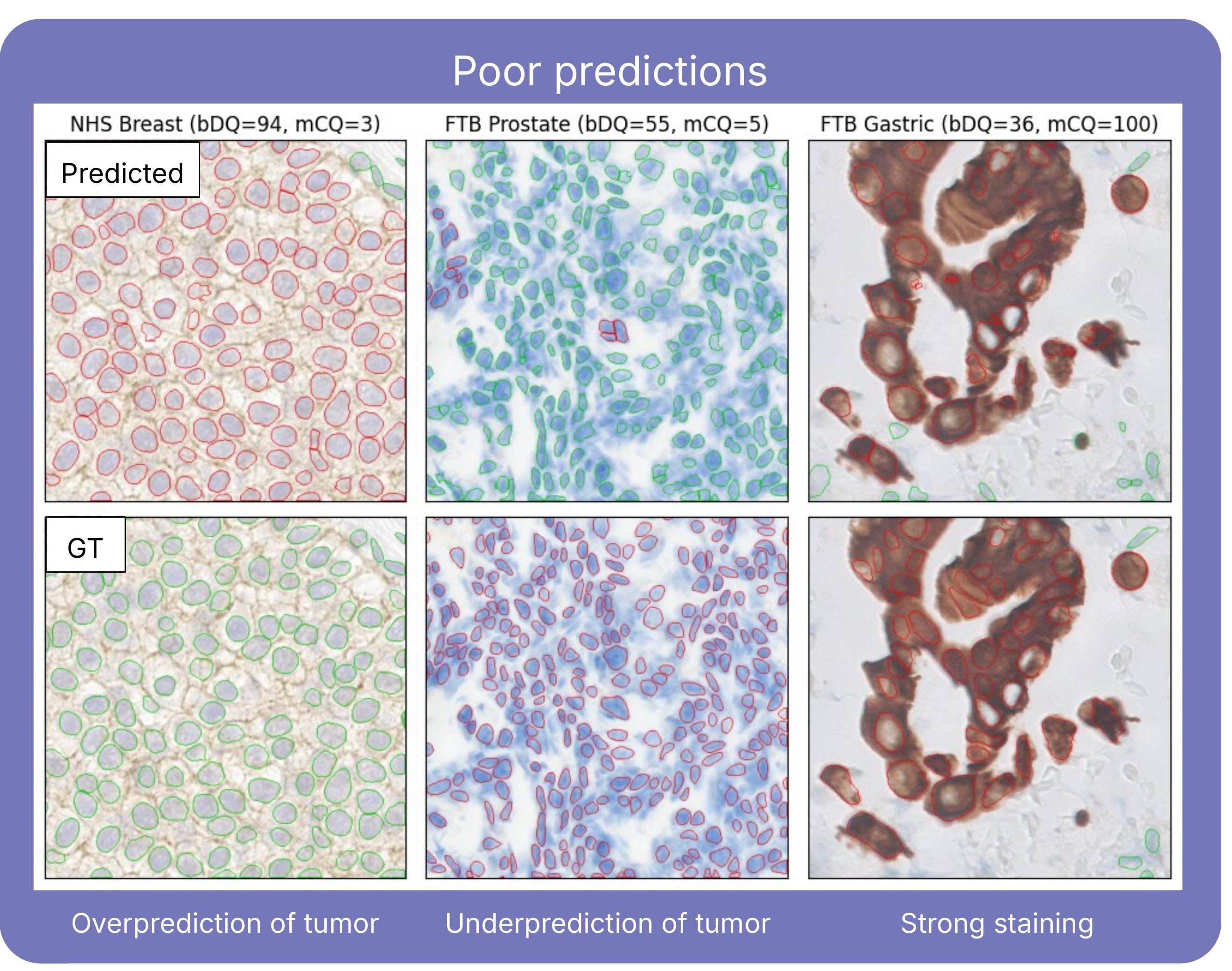
As expected, the primary model performed best on the cohort closest to its training data (breast HER2), and its performance dropped on the three out-of-domain sub-cohorts. Tumor cell classification turned out to be the most sensitive step: it showed the steepest decline when the model encountered a new tumor type, compared with detection and segmentation (Figure 1).
The fine-tuning step is where the results get encouraging. Jointly training with only 20-30 example tiles per indication, performance improved across all three out-of-domain sub-cohorts, suggesting potential in multi-indication and/or multi-marker learning. Tumor cell classification accuracy, the piece that had struggled the most going in, also showed the largest gains after fine-tuning.

Why does tumor-agnostic IHC AI matter for ADC development?
For biopharma teams developing ADCs and other targeted therapies, this kind of groundwork has a direct payoff. New drug programs routinely introduce new biomarkers and new tumor types, and building a bespoke AI model for each one can take months. Few-shot fine-tuning offers a practical way to adapt today, with a small, targeted set of examples rather than a full retraining effort. But the bigger goal is a foundation model trained on a diverse enough mix of indications and markers that it generalizes to new tumor types on its own, with little or no fine-tuning needed. Each new indication added to the training set is a step toward that, as well as being useful in its own right.
This prototype is the first step in the development of a tumor-agnostic AI-based IHC analysis pipeline and testing the approach across more markers, tumor types, and clinical contexts will determine how far it can scale, and that work is already underway.
Developing an ADC or another targeted therapy that needs a new IHC biomarker? Discover our biopharma solutions and talk with the Waiv team about AI-derived biomarkers built for your program.
FAQ
- What does tumor-agnostic mean in IHC AI? It describes a model designed to work across tumor types and IHC markers, so a new indication doesn’t mean building a model from scratch. It isn’t the same as tumor-agnostic therapies, which are approved based on a biomarker rather than where the tumor started.
- Why do ADCs create new IHC scoring needs? Each ADC program can bring a new biomarker, or a new indication for an existing one, and each may need its own scoring criteria. Pathologists have to learn them, and AI tools trained on one marker and tumor type often have to be rebuilt.
- What is few-shot fine-tuning? It means adapting an already trained model to a new tumor type or marker using a small set of annotated examples. Here, the team used 20 to 30 annotated tiles per indication instead of retraining from scratch.
- Is this a diagnostic product? No. It’s a research prototype presented as a conference poster, a first step toward a tumor-agnostic IHC analysis pipeline.
Authors

References
N. A. Koohbanani, “NuClick: A Deep Learning Framework for Interactive Segmentation of Microscopic Images,” Medical Image Analysis, Oct. 2020.
F. Hörst et al., “CellViT: Vision Transformers for precise cell segmentation and classification,” Medical Image Analysis, May 2024.